
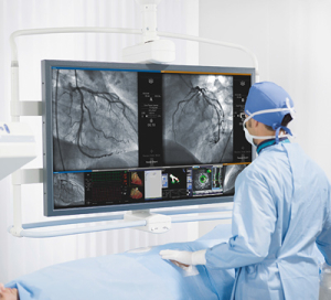
Fig. 1 – Coronary Angiogram and Angioplasty (PCI) are performed in a Cath Lab
What is a Cath lab?
The Cardiac Catheterization Lab (Cath Lab Fig. 1) is a specialized operating suite, where diagnostic and interventional cardiology procedures are performed using small catheters inserted into the heart under X-ray guidance.
What are some common Cath Lab procedures?
- Left Heart Catheterization with Coronary Angiogram
- Right Heart Catheterization
- Coronary Angioplasty (PCI/Stent)
- Pacemaker Device Implantation and Cardiac EP Procedures
- Peripheral Angiogram and Angioplasty
How is Cardiac Catheterization performed?
Tiny catheters are inserted into blood vessels in the wrist or groin (Fig. 2). These catheters are advanced (under X-ray guidance) into the left and right cardiac chambers and the coronary arteries where pressures and oxygen content can be measured. Contrast can then be injected whilst X-rays are recorded to visualize the cardiac chambers or coronary arteries (Fig. 3).
Left heart catheterization provides access to and diagnostic information about the left ventricle, aorta and coronary arteries as well as the aortic and mitral valves. Some interventional procedures such as coronary angioplasty with stenting are performed via catheters during left heart catheterization.
Right heart catheterization provides access to and information about the pulmonary arteries, right ventricle and right atrium. The catheter can be directed across the atrial septum from the right to the left atrium into pulmonary veins or across the mitral valve into the left ventricle and aorta. Pacemaker devices connect to the heart via catheters and leads placed during right heart catheterization.
What is a Coronary Angiogram?
The most common left heart catheterization procedure performed in the Cath Lab is a coronary angiogram. This is an X-ray picture of the heart’s coronary artery, taken when contrast dye is injected into the coronary artery. The angiogram demonstrates whether the artery is normal or narrowed by coronary artery disease. If there are severe blockages found during the cardiac catheterization procedure, your cardiologist may recommend coronary angioplasty (PCI) which is also performed during left heart catheterization. Severe blockages may sometimes require cardiac surgery (open-heart surgery) whereas mild blockages can be treated with medical therapy.
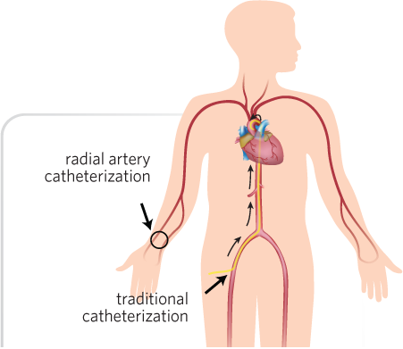
Fig 2 – Tiny catheters are inserted through arteries in the wrist or groin and advanced to the heart.
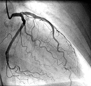
Fig. 3 – Angiogram showing normal coronary arteries visualized with contrast dye during X-ray recording.

Learn more: